Emergent Performance Lab

Share this post
Individual Variations In Skeletal Muscle Vasodilator Capacity: A Case Study
emergentperformance.substack.com

Individual Variations In Skeletal Muscle Vasodilator Capacity: A Case Study

Evan Peikon
Aug 15, 2021
Comment2
Share

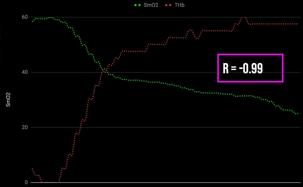
There are local feedback mechanisms in exercising muscles that respond to imbalances in oxygen supply and demand, and adjust vascular conductance which is the ease with which blood flows. This means that when muscle blood flow is compromised during exercise as is often the case during sustained high force muscle contractions, and muscle oxygen saturation levels plummet, there will be a vasodilatory response that works to restore muscle blood flow.  We can observe this in the figure below, which depicts the linear relationship between muscle oxygen saturation and blood volume. For every unit of oxygen consumed, there is a proportionate compensatory increase in muscle blood flow.

Figure I - Muscle oxygen saturation % (green) and [log] total hemoglobin g/dl (red) are graphed against time. There is an inverse linear correlation between these metrics, indicating a proportionate increase in muscle blood volume per unit of deoxygenation.

However, while this response occurs under ideal scenarios there are individuals in which this response is absent. I'm not talking about scenarios where an athlete is contracting a muscle so hard that they restrict blood flow, or scenarios where someone is exercising with so much skeletal muscle mass that the sympathetic nervous system 'protects' their cardiac output by vasoconstricting a tissue. These are normal physiologic scenarios that occur in all individuals. I'm talking about individuals who truly lack the ability to vasodilate in response to deoxygenation, which is associated with a 'non-compensator phenotype'. Individuals with this non-compensator trait experience much greater reductions in performance during high intensity exercise due to an impaired ability to increase muscle blood flow.

The existence of the non-compensator phenotype also speaks to the importance of understanding interindividual differences in the mechanisms that govern oxygen supply and demand relationships. These mechanisms include changes in K+, osmolarity, PO2 and adenosine, which all play a role in the initiation of exercise induced increases in blood flow. However, these factors lack any sustained influence as exercise continues, and most of them are cleared from the active working muscles within minutes of the start of exercise. It appears that sustained vasodilation, and compensatory vasodilation in response to deoxygenation, are more closely related to oxygen sensing by the red blood cells. 

The red blood cell oxygen sensor hypothesis has gained a lot of recognition as a potential  mechanism for matching oxygen supply to oxygen demand. As an individual increases exercise intensity and red blood cell's oxygen saturation decreases, there will be a conformational change in hemoglobin's structure which results in the release of nitric oxide, thus evoking local vasodilation and aiding in the supply of oxygenated blood to the working muscle.  It's speculated that the lack of compensatory vasodilation in athletes with the non-compensators phenotype might indicate compromised red blood cell nitric oxide release in response to oxygen utilization. To give you an idea of what this looks like in practice, in figure II we have NIRS trends from two athletes performing a progressive step test consisting of four minute work bouts at 12.5%, 15%, 17.5%, 20%, 22.5%, 25%, 27.5%, 30%, and 32.5% of their respective maximum sprint speeds with one minute rest between each set. The individuals data who is displayed on top is able to increase skeletal muscle blood flow in proportion to deoxygenation and the individual whose data is displayed on bottom is incapable of doing so.

Figure II. Top - muscle oxygen saturation % (green) and total hemoglobin (red) for the compensator phenotype athlete. Bottom - - muscle oxygen saturation % (green) and total hemoglobin (red) for the non-compensator phenotype athlete.

The differences in the individuals with the compensator versus non-compensator phenotype are also present in the respective correlations between muscle oxygen saturation and total hemoglobin for these two individuals. For the athletes with the compensator phenotype (top) the R value ranged from -0.94 to -0.98 for all work bouts whereas the athlete with the non-compensator phenotype (bottom) has R values ranging from -0.44 to -0.7 on all work bouts. The latter individual also reported much higher RPE’s on each workout, and they presented with both a greater +ΔHR and +ΔRPE from bout to bout. In practice i’ve also found the latter athlete who have much more compromised work capacity when they exercise at intensities above their critical power in the 'severe' work domain.

The question now becomes how we improve the non-compensators ability to vasodilate over time. This is an ongoing area of investigation for me and my colleagues, and I suspect we'll have some more conclusive answers as time goes on. As of now, it appears that the best bet is to select training modalities that improve peripheral circulation and mitochondrial density, increase dietary nitrate consumption (beets), and address basic lifestyle factors related to blood sugar control, vascular health, and tissue circulation. Below i’ve posted a sample two week snapshot from this athletes training, roughly eight months into our time working together as they prep for special forces selection.

Figure III. Two week training program (A week, B week) from an individual prepping for special forces section. All training sessions are based on their ‘sport specific’ limitations, physiological limitations, are personal training goals.

In addition to seeing performance improvements in all of the major variables we sought to train, we were also able to improve this individuals vasodilator capacity. In figure IV below we have their ΔTHb trends from before and after a multi week training intervention where they completed a 60 second Echo Bike @40% MSS with 2:00 rest between sets. Notice the difference in ΔTHb responses during the recovery periods before and after the intervention. This indicates an improved vasodilator capacity and an increased hyperemic response to exercise.

Figure IV - An individuals reactive hyperemia response to a 60x5 second Echo Bike @40% MS with 2:00 rest between intervals pre and post training intervention

If you enjoyed reading this article please consider subscribing to the Emergent Performance Lab Substack. 

  • Free subscribers can expect content short form posts sporadically throughout the month, as well as previews of my long form paid content. 

  • Paid subscribers will receive 1-2 content pieces per week including one long form exclusive content piece every Sunday, as well as bonus content including mini-articles, case studies, monthly Q&A’s, and podcast.

Comment2
ShareShare

Create your profile

0 subscriptions will be displayed on your profile (edit)

Skip for now

Only paid subscribers can comment on this post

Already a paid subscriber? Sign in

Check your email

For your security, we need to re-authenticate you.

Click the link we sent to , or click here to sign in.

Brian Heep
Aug 15, 2021Liked by Evan Peikon

Evan, subscribed about a month ago- little more, and have gone back and read your other posts. As an owner of the PNOE metabolic device, heard you on Daniels podcast. Really enjoy the content you're putting out and answering the questions we've all had of why (myself or others I'm working with) simply give out at certain intensities and just the variability within individual workload capacities. Thought this post broke that down really well. I appreciate it.

Expand full comment
Reply
1 reply by Evan Peikon
1 more comments…
TopNewCommunity

No posts

Ready for more?

© 2022 Evan Peikon
Privacy ∙ Terms ∙ Collection notice
Publish on Substack Get the app
Substack is the home for great writing