

Why Does My Lower Back Always Blow Up On Deadlifts?
Why Does My Lower Back Always Blow Up On Deadlifts?
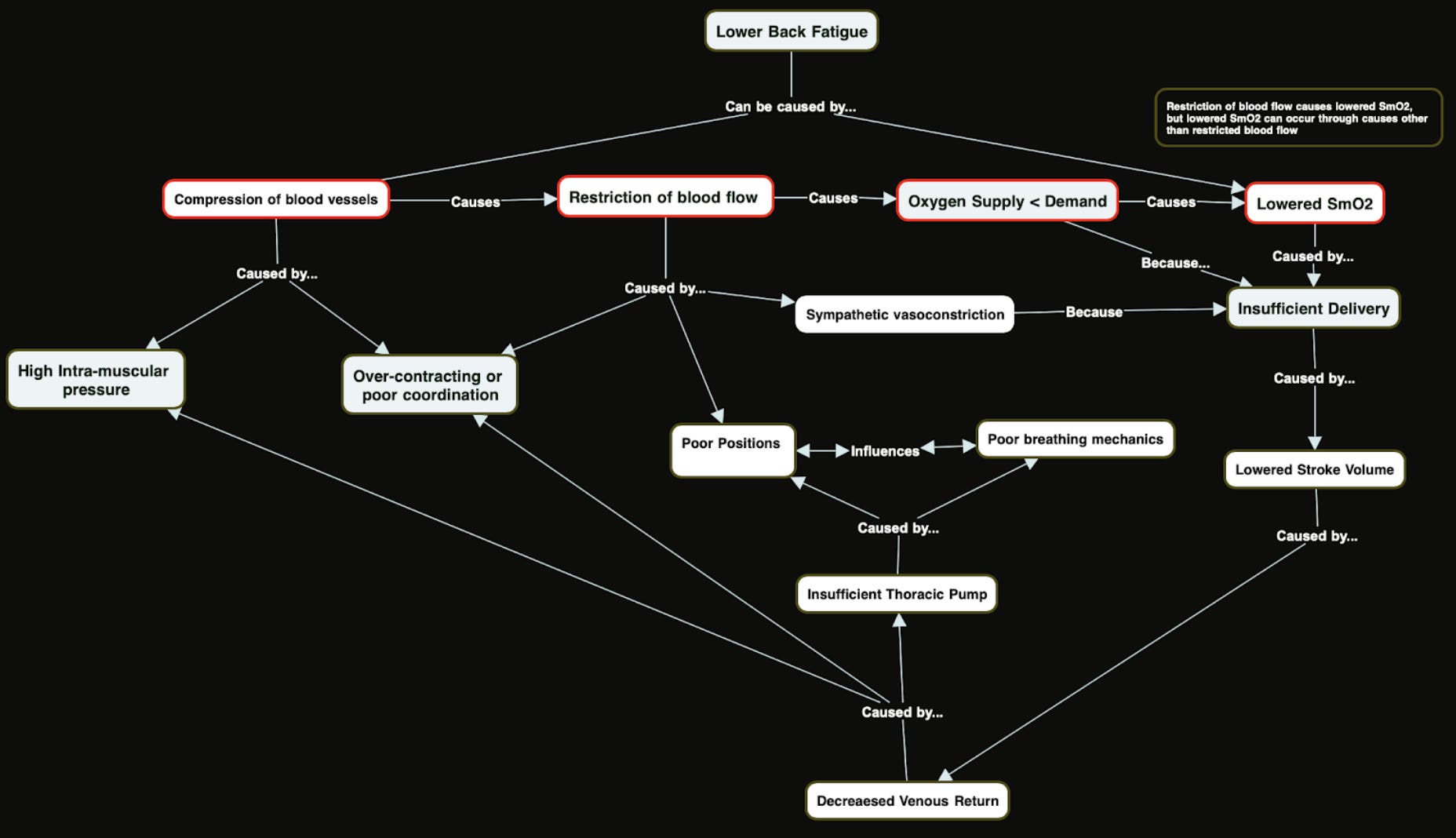
A Systems Approach

As a sports scientist, one of things I love getting to do is taking concepts from research and applying them in practice to see what the outcome is for an individual. If there is a specific problem i’m trying to solve i’ll comb the research for some solutions, then put them to the test and observe the results. Sometimes clear trends emerge through this process and these protocols trickle down into my coaching practice, and other times the observed responses are so variable between individuals that it’s difficult to parse out why something did or didn’t get the desired effect.
Recently i’ve been interested in seeing why some athletes’ lower backs always ‘blow up’ during metcons (even metcons that don’t have a meaningful hinging component) while other athletes almost never experience this. I’ve observed a trend that the Crossfit athletes who have these lower back issues tend to be stronger and more heavily muscle individuals, and often times they’re also the athletes with weak cardiac ou…
Keep reading with a 7-day free trial
Subscribe to On Human Performance by Evan Peikon to keep reading this post and get 7 days of free access to the full post archives.